7 Levels of Lymph Nodes in Neck: A Comprehensive Guide to Anatomy and Function
The human neck is a complex region, home to vital structures like blood vessels, nerves, and the lymphatic system. Among these, the lymph nodes play a critical role in immunity, filtering harmful substances and aiding in the fight against infection and disease. When it comes to the neck, medical professionals classify these nodes into 7 levels of lymph nodes in neck, a system that’s essential for diagnosing and treating conditions like infections, inflammation, and cancer. As of March 12, 2025, this guide explores the anatomy, function, and clinical significance of these seven levels, offering a detailed resource for students, healthcare providers, and curious minds alike.

What Are Lymph Nodes and Why Do They Matter?
Lymph nodes are small, bean-shaped glands scattered throughout the body, with about 300 of the body’s 800 nodes located in the neck. They’re part of the lymphatic system, which filters lymph fluid—a mix of waste, fluid, and immune cells—before returning it to the bloodstream. In the neck, these nodes act as sentinels, catching pathogens or cancer cells from the head and upper respiratory tract. The 7 levels of lymph nodes in neck provide a standardized map for identifying their location and drainage patterns, crucial for medical imaging, surgery, and cancer staging.
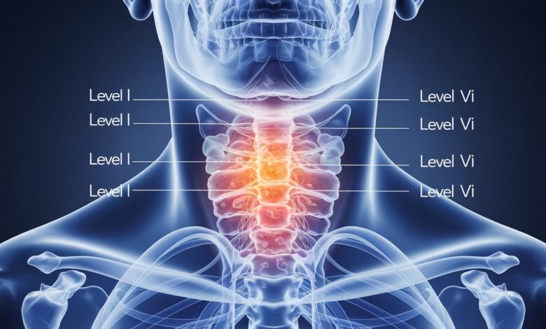
This classification, developed by the American Academy of Otolaryngology-Head and Neck Surgery and refined over decades, divides the neck’s lymph nodes into seven distinct zones (Levels I-VII). Each level corresponds to specific anatomical boundaries and drainage areas, making it a cornerstone of head and neck medicine. Let’s dive into these levels and uncover their roles.
The 7 Levels of Lymph Nodes in Neck: Anatomy and Drainage
Level I: Submental and Submandibular Nodes
- Location: Level I is split into IA (submental) and IB (submandibular). IA sits between the anterior bellies of the digastric muscles, below the chin, and above the hyoid bone. IB lies between the anterior and posterior digastric bellies, beneath the mandible.
- Drainage: IA filters lymph from the lower lip, floor of the mouth, and anterior tongue. IB drains the oral cavity, cheeks, lips, and submandibular gland.
- Clinical Relevance: Swelling here often signals dental infections or oral cancers. Surgeons may perform a Level I dissection for tongue or lip carcinomas.
Level II: Upper Jugular Nodes
- Location: From the skull base to the hyoid bone, along the upper third of the internal jugular vein. It’s divided into IIA (anterior to the spinal accessory nerve) and IIB (posterior to it).
- Drainage: Receives lymph from the face, parotid gland, nasopharynx, oropharynx, and larynx. The jugulodigastric node, a key player, is often the first to swell in throat infections.
- Clinical Relevance: Level II is a hotspot for metastases from head and neck cancers, especially nasopharyngeal or oropharyngeal tumors. IIB is trickier to access surgically due to the spinal accessory nerve.
Level III: Middle Jugular Nodes
- Location: Extends from the hyoid bone to the cricoid cartilage, along the middle third of the internal jugular vein.
- Drainage: Filters lymph from the base of the tongue, tonsils, larynx, hypopharynx, and thyroid gland.
- Clinical Relevance: Nodes here swell with throat infections like tonsillitis or signal cancers of the larynx or hypopharynx. It’s a common site for staging in head and neck oncology.
Level IV: Lower Jugular Nodes
- Location: From the cricoid cartilage to the clavicle, along the lower third of the internal jugular vein.
- Drainage: Drains the hypopharynx, larynx, thyroid, and cervical esophagus.
- Clinical Relevance: Level IV includes the Virchow node (left side), a sentinel for abdominal cancers like gastric carcinoma. It’s also key in thyroid cancer staging.
Level V: Posterior Triangle Nodes
- Location: In the posterior triangle, bounded by the sternocleidomastoid muscle (anteriorly), trapezius muscle (posteriorly), and clavicle (inferiorly). Split into VA (above the cricoid) and VB (below it).
- Drainage: Collects lymph from the scalp, neck skin, nasopharynx, and occipital region.
- Clinical Relevance: Often involved in skin cancers (e.g., melanoma) or nasopharyngeal carcinoma. Supraclavicular nodes in VB can indicate distant metastases.
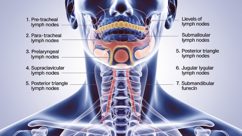
Level VI: Anterior Compartment Nodes
- Location: Central neck, from the hyoid bone to the suprasternal notch, between the carotid sheaths. Includes prelaryngeal, pretracheal, and paratracheal nodes.
- Drainage: Drains the thyroid gland, larynx, trachea, and esophagus.
- Clinical Relevance: Critical in thyroid cancer, where Level VI dissection is routine. The Delphian node (prelaryngeal) is a marker for laryngeal or thyroid malignancy.
Level VII: Superior Mediastinal Nodes
- Location: Below the suprasternal notch, extending into the upper mediastinum to the innominate artery.
- Drainage: Filters lymph from the lower neck, thyroid, trachea, and esophagus.
- Clinical Relevance: Often included in central neck dissection for papillary thyroid cancer. Metastases here upstage the disease to Stage IVa per AJCC guidelines.
How the 7 Levels Were Established
The 7 levels of lymph nodes in neck system evolved from early anatomical studies by Henri Rouvière in 1932, who mapped cervical lymphatics. In the 1930s, Memorial Sloan Kettering Cancer Center formalized a numbered classification, later standardized by the American Academy of Otolaryngology in 1991 and updated in 2002 with sublevels (e.g., IIA/IIB). The American Joint Committee on Cancer (AJCC) adopted it for staging, with the 8th edition (2018) still in use as of 2025. This framework balances surgical landmarks with imaging-based precision, making it indispensable for modern medicine.
Clinical Importance of the 7 Levels
Cancer Staging and Treatment
Head and neck cancers—squamous cell carcinoma, thyroid cancer, lymphoma—spread predictably through these lymph node levels. For example:
- Oral cavity cancers hit Levels I-III.
- Nasopharyngeal cancers target Levels II and V.
- Thyroid cancers involve Levels VI and VII.
Surgeons use selective neck dissections (e.g., Levels II-IV for oropharyngeal cancer) or comprehensive ones based on this map. Radiation oncologists target specific levels to minimize damage to healthy tissue.
Infection and Inflammation
Swollen nodes signal infection (e.g., tonsillitis in Level II) or inflammation (e.g., autoimmune diseases). Their location pinpoints the source—Level IA swelling might mean a lip infection, while Level VI could indicate thyroiditis.
Diagnostic Imaging
Ultrasound, CT, and MRI rely on the 7 levels of lymph nodes in neck to localize suspicious nodes. A rounded, enlarged node in Level IV with extracapsular spread on CT might prompt a biopsy for metastasis.
How to Assess Lymph Nodes in the Neck
- Physical Exam: Palpate each level, feeling for size (>1 cm is abnormal), tenderness, or fixation (cancer nodes are hard and fixed).
- Imaging: Ultrasound detects shape and echogenicity; CT/MRI assess size and necrosis.
- Biopsy: Fine needle aspiration (FNA) or excisional biopsy confirms malignancy if imaging is inconclusive.
Tips for Keeping Lymph Nodes Healthy
- Hydration: Supports lymph flow.
- Diet: Anti-inflammatory foods (e.g., berries, leafy greens) reduce strain on the system.
- Avoid Infection: Good hygiene prevents overload from pathogens.
Common Questions About the 7 Levels
Why 7 Levels and Not More?
The system balances detail with practicality. Other nodes (e.g., parotid, occipital) exist but aren’t routinely dissected, so they’re excluded from the core seven.
Can Swollen Nodes Be Normal?
Yes, reactive swelling from infection is common and resolves post-recovery. Persistent or painless enlargement warrants investigation.
How Do Surgeons Access Level VII?
Through a cervical incision, extending into the mediastinum if needed—safe but requires precision to avoid nerve injury.
Comparing the Levels
| Level | Location | Drainage Area | Common Issues |
|---|---|---|---|
| I | Submental/Submandibular | Mouth, lips, tongue | Oral infections, cancers |
| II | Upper jugular | Face, throat, parotid | Throat cancers, tonsillitis |
| III | Middle jugular | Tongue, larynx, thyroid | Laryngeal cancer |
| IV | Lower jugular | Larynx, esophagus, thyroid | Thyroid cancer, metastases |
| V | Posterior triangle | Scalp, neck skin, nasopharynx | Skin cancer, lymphoma |
| VI | Anterior compartment | Thyroid, trachea, larynx | Thyroid cancer |
| VII | Superior mediastinum | Lower neck, esophagus | Advanced thyroid cancer |
Conclusion
The 7 levels of lymph nodes in neck—from Level I under the chin to Level VII in the mediastinum—form a vital framework for understanding neck anatomy and managing health conditions. As of March 12, 2025, this system remains the gold standard in oncology, surgery, and diagnostics, guiding everything from cancer staging to infection tracking. Each level’s unique drainage pattern and clinical role make it a map of the body’s immune defenses in the neck. Whether you’re a medical professional staging a tumor or a patient curious about a swollen gland, grasping these seven levels empowers you to navigate health with confidence. Keep this guide handy, and you’ll never see your neck the same way again!